Radiographic Analysis (X-Ray)
Forensic identification of anomalous shape in extraction cavity.

Technical Metadata (DICOM)
| Source Format | DICOM (Medical Standard) |
| Export Method | Lossless 16-bit PNG |
| Sensor Type | Intraoral Digital Sensor |
| Capture Date | Feb 15, 2022 | 13:49:30 |
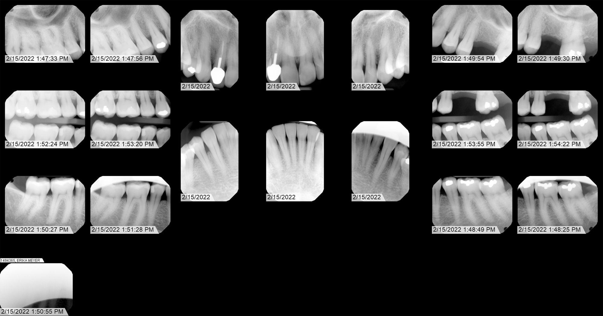
Case Narrative: Tooth 14 Anomaly
This investigation focuses on a localized, distinctly anomalous artifact identified in the jaw area, embedded in the jawbone over an extraction cavity (Tooth 14). The geometric angles and density of this finding suggest a non-biological origin.
The significance of this radiograph is further reinforced by a history of institutional resistance encountered during attempts at verification. Despite significant effort throughout 2022, I was unable to obtain original DICOM files from either Advantage Dental or Roots Dental. However, in 2026 I was able to locate and cite the federal and state laws requiring them to provide me with that information: HIPAA (45 CFR § 164.524) and Oregon Administrative Rule (OAR) 818-012-0030. As of this writing, Advantage Dental has provided me with those files, and to their credit, they did so quickly. I am still waiting for the single matching radiograph in DICOM format from Roots Dental, taken on April 8, 2022. Once I have that information, I'll create a page comparing the two.
Under the principles established by the 21st Century Cures Act and SWGDE forensic standards, original DICOM data is considered the "Primary Evidence." Prior to this year, these dentists insisted on substituting lower-resolution exports, effectively preventing the authentication of sensor-level metadata, a practice recognized in forensic circles as a barrier to independent verification.
Secondary Exhibits


Dental map showing numbered teeth. Tooth 14 extracted Jan 10, 2020